Bone biology
Understanding bone biology
Bone is not an inert scaffold. It is living, vascular, mineralized tissue that senses load, repairs microdamage, stores minerals, houses marrow, and continuously rebuilds itself through coordinated cellular and molecular signaling.
The big picture: bone from organ to molecule
Bone biology can be understood in layers. At the gross level, a long bone contains cortical bone, trabecular bone, marrow, periosteum, endosteum, blood vessels, and nerves. At the microscopic level, compact bone is organized into osteons, lamellae, lacunae, and canaliculi. At the molecular level, collagen, hydroxyapatite, RANK/RANKL/OPG, WNT signaling, sclerostin, cytokines, and hormones help determine whether bone is being built, maintained, or resorbed.12

Gross structure: cortical bone, trabecular bone, marrow, and periosteum
Cortical bone is the dense outer shell that gives long bones much of their bending and torsional strength. Trabecular, or cancellous, bone forms an internal lattice that is metabolically active and responsive to loading. The periosteum covers the outer bone surface and contains nerves, vessels, and progenitor cells; the endosteum lines inner surfaces where remodeling is also active.12

Microstructure: osteons, lamellae, lacunae, and canaliculi
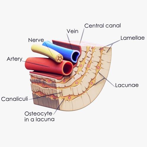
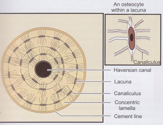
An osteon, also called a Haversian system, is a cylindrical structural unit of cortical bone. Concentric lamellae are rings of mineralized matrix surrounding a central canal. Osteocytes sit in lacunae between lamellae and extend processes through canaliculi, allowing cell-to-cell communication and fluid-flow sensing.134

The matrix: collagen plus mineral, not just calcium
Bone matrix begins as osteoid, an organic matrix made mainly of type I collagen. Mineralization then deposits hydroxyapatite crystals into and around that collagen scaffold. Collagen gives bone toughness and resistance to cracking; mineral gives stiffness and compressive strength.111
Bone remodeling: how bone renews itself
Adult bone is renewed by basic multicellular units. Osteoclasts resorb older or damaged bone, reversal cells help prepare the surface, and osteoblasts refill the cavity with new osteoid that later mineralizes. This remodeling process repairs microdamage and helps calcium-phosphate homeostasis, but it can weaken bone if resorption repeatedly outpaces formation.25
Bone cells and what they do
| Cell | Origin / location | Main function | Important signals |
|---|---|---|---|
| Osteoprogenitor / mesenchymal stromal cell | Periosteum, endosteum, marrow stromal compartment | Precursor pool for osteoblast-lineage cells; responds to mechanical and hormonal signals. | RUNX2, SP7/osterix, BMPs, WNT, PTH/PTH1R |
| Osteoblast | Bone-forming surface cell from mesenchymal lineage | Produces osteoid, type I collagen, alkaline phosphatase, and mineralization-regulating proteins. | WNT/beta-catenin, RUNX2, SP7, BMPs, ALPL, BGLAP, SPP1 |
| Osteocyte | Former osteoblast embedded in lacunae | Most abundant mature bone cell; senses mechanical load, regulates mineral metabolism, and coordinates remodeling. | SOST/sclerostin, RANKL, FGF23, connexins, nitric oxide, prostaglandins |
| Bone lining cell | Flattened quiescent osteoblast-lineage cell on bone surfaces | Covers resting surfaces; can help regulate access to mineralized surface and participate in remodeling activation. | RANKL/OPG balance, PTH response, local coupling signals |
| Osteoclast precursor | Hematopoietic monocyte-macrophage lineage | Fuses into multinucleated osteoclasts when exposed to key differentiation signals. | M-CSF, RANK, RANKL, NF-kB, NFATc1 |
| Osteoclast | Multinucleated hematopoietic-lineage cell on bone surface | Resorbs bone by acidifying the sealed resorption zone and degrading matrix. | RANKL, integrins, cathepsin K, TRAP, calcitonin receptor |
| Chondrocyte | Cartilage cell, especially growth plate and fracture callus contexts | Builds cartilage matrix; participates in endochondral ossification during growth and repair. | SOX9, Indian hedgehog, PTHrP, collagen type II and X |
| Immune and marrow cells | Bone marrow | Influence remodeling through inflammatory cytokines and marrow-bone crosstalk. | IL-1, IL-6, TNF-alpha, interferons, RANKL |
RANK, RANKL, and osteoprotegerin: the osteoclast control system
RANKL is a key signal made by osteoblast-lineage cells and osteocytes. It binds RANK on osteoclast precursors and activates downstream pathways that drive osteoclast differentiation, activation, and survival. Osteoprotegerin, abbreviated OPG, is a soluble decoy receptor that binds RANKL and prevents it from activating RANK.67
Cell-level detail: RANK signaling from membrane to nucleus to matrix resorption
Key: RANKL = receptor activator of nuclear factor kappa-B ligand (TNFSF11 protein/gene); RANK = receptor activator of nuclear factor kappa-B (TNFRSF11A receptor/gene); OPG = osteoprotegerin, a soluble RANKL decoy receptor (TNFRSF11B); M-CSF = macrophage colony-stimulating factor, also called CSF1 (CSF1 protein/gene); c-Fms = M-CSF receptor (CSF1R protein/gene); TRAF6 = TNF receptor-associated factor 6 adapter; IKK = IkB kinase complex; NF-kB = nuclear factor kappa-B transcription-factor family; MAPK = mitogen-activated protein kinase pathway; AP-1 = activator protein-1 transcription-factor complex; c-Fos = FOS protein/gene component of AP-1; Ca2+ = calcium signal; calcineurin = calcium-dependent phosphatase; NFATc1 = nuclear factor of activated T-cells c1 (NFATC1 protein/gene); CTSK = cathepsin K; ACP5/TRAP = tartrate-resistant acid phosphatase; DCSTAMP = dendritic cell-specific transmembrane protein; ATP6V0D2 = vacuolar ATPase V0 subunit d2; ITGB3 = beta-3 integrin; TCIRG1 = vacuolar proton-pump a3 subunit; V-ATPase/H+ = vacuolar ATPase proton pump; ruffled border and actin sealing zone = osteoclast resorption structures; type I collagen = COL1A1/COL1A2 collagen matrix.
WNT, LRP5/6, beta-catenin, and sclerostin: the bone-formation gate
Canonical WNT signaling helps osteoblast-lineage cells commit to bone formation and supports osteoblast function. WNT ligands signal through Frizzled receptors and LRP5/6 coreceptors, allowing beta-catenin to accumulate and activate bone-forming gene programs. Sclerostin, encoded by the SOST gene and produced mainly by osteocytes, inhibits LRP5/6 and restrains bone formation.89
Cell-level detail: WNT signaling from matrix environment to nuclear bone-formation genes
That is why osteocytes are so important: they do not simply sit inside bone. They help decide whether the local skeleton should build, conserve, or remodel. Mechanical loading and intermittent parathyroid hormone signaling tend to reduce sclerostin, while unloading tends to favor signals that reduce formation.3810
Molecules, proteins, collagens, cytokines, and pathways
| Molecule / pathway | Category | Plain-English role in bone |
|---|---|---|
| Type I collagen (COL1A1/COL1A2) | Structural matrix protein | Main organic scaffold of bone; contributes toughness and crack resistance. |
| Hydroxyapatite | Mineral crystal | Calcium-phosphate crystal that stiffens the collagen matrix. |
| Alkaline phosphatase (ALPL) | Mineralization enzyme / formation marker | Supports mineralization; bone-specific alkaline phosphatase is a formation marker. |
| Osteocalcin (BGLAP) | Noncollagen bone matrix protein | Osteoblast product associated with mineralized matrix and bone formation. |
| Osteopontin (SPP1) | Matrix glycoprotein | Helps regulate cell attachment and mineral-matrix interactions. |
| Osteonectin / SPARC | Matrix glycoprotein | Binds collagen and mineral; helps organize mineralized matrix. |
| RANKL (TNFSF11) | Osteoclast differentiation cytokine | Key “go” signal for osteoclast formation and survival. |
| RANK (TNFRSF11A) | Receptor | Receptor on osteoclast precursors that receives the RANKL signal. |
| Osteoprotegerin / OPG (TNFRSF11B) | Decoy receptor | Binds RANKL before it reaches RANK, reducing osteoclast signaling. |
| M-CSF (CSF1) | Growth factor | Supports osteoclast precursor survival and differentiation. |
| NF-kB and NFATc1 | Transcription signaling | Downstream RANK signals that help drive osteoclast gene programs. |
| WNT ligands | Formation pathway signal | Promote osteoblast-lineage commitment and bone formation signaling. |
| LRP5/6 and Frizzled | WNT receptor complex | Receives WNT signals at the cell surface. |
| Beta-catenin (CTNNB1) | Canonical WNT effector | Moves into the nucleus when WNT signaling is active and supports bone-forming gene expression. |
| Sclerostin (SOST) | Osteocyte-derived WNT inhibitor | Acts as a brake on bone formation by inhibiting LRP5/6. |
| DKK1 | WNT inhibitor | Another inhibitor of LRP5/6-mediated WNT signaling. |
| RUNX2 and SP7/osterix | Osteoblast transcription factors | Help immature cells commit to the osteoblast lineage. |
| BMPs | Growth factors | Promote osteoblast differentiation and matrix production in many contexts. |
| TGF-beta | Growth factor released from matrix | Participates in coupling resorption to formation and cell recruitment. |
| IL-1, IL-6, TNF-alpha | Inflammatory cytokines | Can increase resorption signaling in inflammatory states. |
| PTH / PTH1R | Hormone and receptor | Regulates calcium and remodeling; intermittent and continuous exposure have different skeletal effects. |
| FGF23 | Osteocyte hormone | Helps regulate phosphate and vitamin D metabolism through kidney signaling. |
| PINP, CTX, TRAP, DPD | Bone turnover markers | Blood or urine markers that reflect formation or resorption activity, not structure by themselves. |
This table is a teaching map, not a complete molecular atlas. Bone biology involves many additional genes, receptors, enzymes, and local signals.181112
How to read bone biology clinically
Bone density tests estimate mineral content, but a living skeleton is more complicated than a number. A useful clinical view asks about architecture, turnover, nutrition, hormones, kidney function, medications, inflammation, muscle, falls, and prior fracture history. The molecular pathways above are not meant to make the page a treatment guide; they explain why bone can change over time and why different bone conditions may need different conversations.
Keep learning
About this page
This page is educational and is not medical advice. It is meant to help patients and interested readers understand the basic structure, cells, and molecular pathways of bone before discussing personal risk, testing, or treatment decisions with a clinician.
References
- Florencio-Silva R, Sasso GR, Sasso-Cerri E, Simoes MJ, Cerri PS. Biology of Bone Tissue: Structure, Function, and Factors That Influence Bone Cells. Biomed Res Int. 2015;2015:421746. doi:10.1155/2015/421746. PMID: 26247020. PubMed
- Siddiqui JA, Partridge NC. Physiological Bone Remodeling: Systemic Regulation and Growth Factor Involvement. Physiology (Bethesda). 2016;31(3):233-245. doi:10.1152/physiol.00061.2014. PMID: 27053737. PubMed
- Robling AG, Bonewald LF. The Osteocyte: New Insights. Annu Rev Physiol. 2020;82:485-506. doi:10.1146/annurev-physiol-021119-034332. PMID: 32040934. PubMed
- Schaffler MB, Cheung WY, Majeska R, Kennedy O. Osteocytes: master orchestrators of bone. Calcif Tissue Int. 2014;94(1):5-24. doi:10.1007/s00223-013-9790-y. PMID: 24042263. PubMed
- Delaisse JM. The reversal phase of the bone-remodeling cycle: cellular prerequisites for coupling resorption and formation. Bonekey Rep. 2014;3:561. doi:10.1038/bonekey.2014.56. PMID: 25120911. PubMed
- Boyce BF, Xing L. Functions of RANKL/RANK/OPG in bone modeling and remodeling. Arch Biochem Biophys. 2008;473(2):139-146. doi:10.1016/j.abb.2008.03.018. PMID: 18395508. PubMed
- Boyle WJ, Simonet WS, Lacey DL. Osteoclast differentiation and activation. Nature. 2003;423(6937):337-342. doi:10.1038/nature01658. PMID: 12748652. PubMed
- Baron R, Kneissel M. WNT signaling in bone homeostasis and disease: from human mutations to treatments. Nat Med. 2013;19(2):179-192. doi:10.1038/nm.3074. PMID: 23389618. PubMed
- Glass DA 2nd, Bialek P, Ahn JD, et al. Canonical Wnt signaling in differentiated osteoblasts controls osteoclast differentiation. Dev Cell. 2005;8(5):751-764. doi:10.1016/j.devcel.2005.02.017. PMID: 15866165. PubMed
- Dallas SL, Prideaux M, Bonewald LF. The osteocyte: an endocrine cell … and more. Endocr Rev. 2013;34(5):658-690. doi:10.1210/er.2012-1026. PMID: 23612223. PubMed
- Seibel MJ. Biochemical markers of bone turnover: part I: biochemistry and variability. Clin Biochem Rev. 2005;26(4):97-122. PMID: 16648882. PubMed
- Garnero P. The Utility of Biomarkers in Osteoporosis Management. Mol Diagn Ther. 2017;21(4):401-418. doi:10.1007/s40291-017-0272-1. PMID: 28271451. PubMed
- Gohda J, Akiyama T, Koga T, Takayanagi H, Tanaka S, Inoue J. RANK-mediated amplification of TRAF6 signaling leads to NFATc1 induction during osteoclastogenesis. EMBO J. 2005;24(4):790-799. doi:10.1038/sj.emboj.7600564. PMID: 15678102. PubMed
- Vaananen HK, Zhao H, Mulari M, Halleen JM. The cell biology of osteoclast function. J Cell Sci. 2000;113(Pt 3):377-381. PMID: 10639325. PubMed
- Krishnan V, Bryant HU, MacDougald OA. Regulation of bone mass by Wnt signaling. J Clin Invest. 2006;116(5):1202-1209. doi:10.1172/JCI28551. PMID: 16670761. PubMed
- Li X, Zhang Y, Kang H, Liu W, Liu P, Zhang J, Harris SE, Wu D. Sclerostin binds to LRP5/6 and antagonizes canonical Wnt signaling. J Biol Chem. 2005;280(20):19883-19887. doi:10.1074/jbc.M413274200. PMID: 15778503. PubMed