
Real radiograph source library
Fragility Fracture Radiograph Library
A leereichelMD.com fracture image library built from real radiographs in open-access clinical research articles. The focus is fragility-type fracture patterns: wrist, proximal humerus, distal femur, pelvis, and femoral neck.
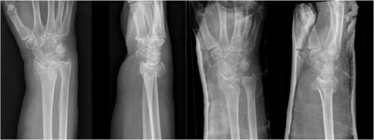
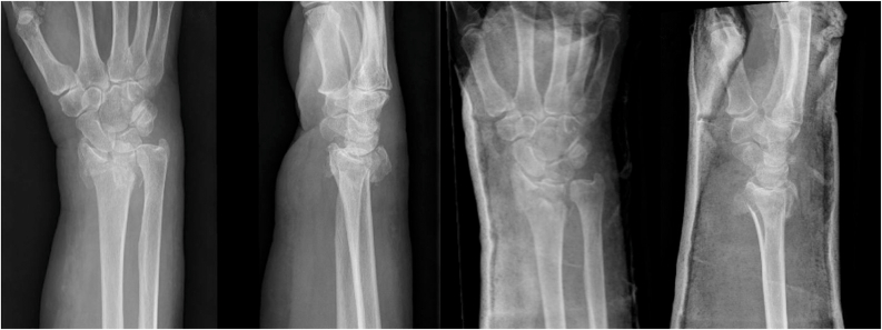
Distal radius / Colles-type fragility pattern
Wrist Fracture
Fragility-type basis: The source study used radiographs and elderly patient profiles for displaced distal radius fractures; this is a common sentinel fragility-fracture pattern in older adults.1
Source: Figure 1 from Burgert et al., CC BY. Cropped status: no crop; original radiograph strip preserved. PMC13217588.
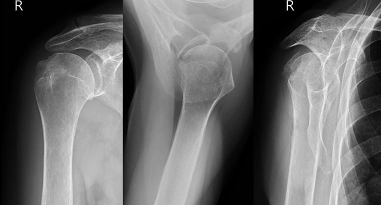
Low-energy elderly shoulder fracture pattern
Proximal Humerus Fracture
Fragility-type basis: The source article frames proximal humerus fractures in older adults as commonly related to standing-height falls, osteoporosis, and low-energy mechanisms.2
Source: Figure 3 from Kalacun et al., CC BY. Cropped status: no crop; original radiograph strip preserved. PMC12532694.
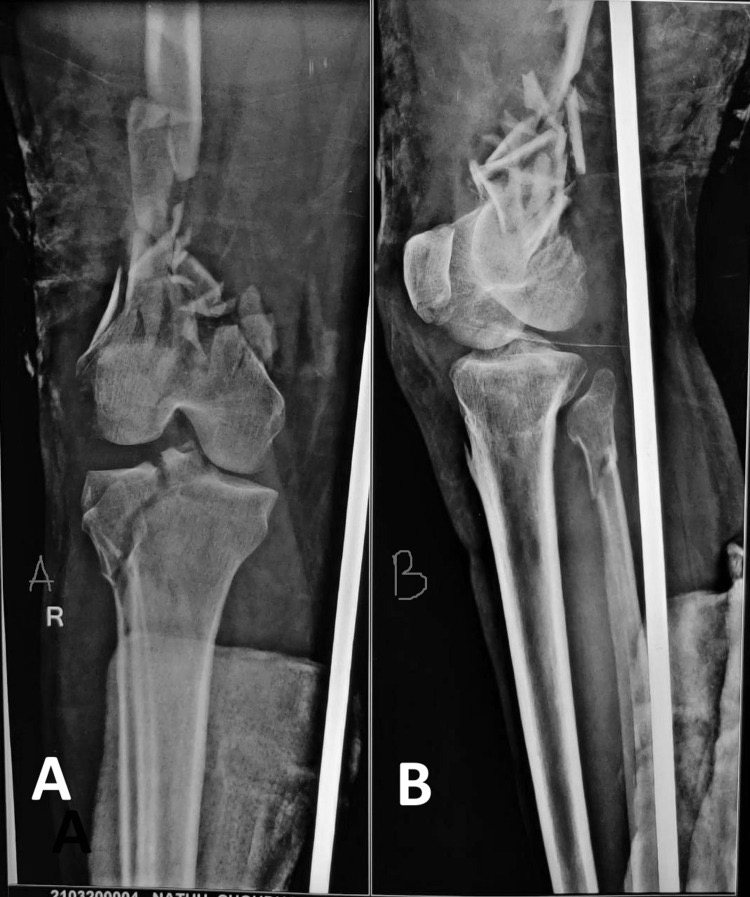
Osteoporotic low-energy distal femur fracture
Distal Femur Fracture
Fragility-type basis: The source cohort was predominantly older and low-energy: the authors reported low-energy mechanisms in most cases, with the age 65-and-older subgroup entirely low-energy.3
Source: Figure 1 from Joshi et al., CC BY. Change made: grayscale normalization from the published film-photo color cast. PMC13080954.
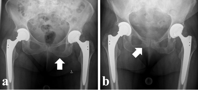
Pubic ramus and pelvic insufficiency fracture pattern
Pelvic Fracture
Fragility-type basis: The source case describes chronic pelvic insufficiency fractures after a lateral fall and highlights the crescendo pattern of osteoporotic pelvic fractures.4
Source: Figure 1 panels a-b from Gewiess et al., CC BY. Change made: cropped to radiograph panels only. PMC11663159.
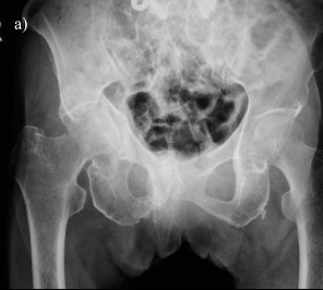
Elderly hip fracture / fragility-fracture context
Femoral Neck Fracture
Fragility-type basis: The source case series concerns elderly fragility fractures of the pelvis with acetabular and femoral neck fractures, including cases without a clear trauma history.5
Source: Figure 1 panel a from Nakai and Fukunishi, CC BY. Change made: cropped to the preoperative radiograph panel only. PMC12262157.
References
- Burgert N, Ter Meulen DP, Schep NWL, Colaris JW, Jonkers HAF, Willigenburg NW, et al. Surgeon preferences and outcome predictions for displaced distal radius fractures in elderly patients: a survey of 151 surgeons making 4,832 treatment predictions based on patient profiles and radiographs. J Hand Surg Glob Online. 2026;8(4):101030. doi:10.1016/j.jhsg.2026.101030. PMID: 42220440. PMCID: PMC13217588.
- Kalacun D, Komadina R, Brilej D. Proximal humerus fractures in the elderly work up, classifications and fracture biomechanics. Eur J Trauma Emerg Surg. 2025;51(1):300. doi:10.1007/s00068-025-02945-0. PMID: 41099833. PMCID: PMC12532694.
- Joshi N, Sharma D, Deshkmukh S, Mankar S, Agrawal PP, Zodey K, et al. Functional and radiological outcomes of distal femur fractures treated with a combination of supracondylar nails and lateral condylar locking plates. Cureus. 2026;18(3):e105075. doi:10.7759/cureus.105075. PMID: 41994725. PMCID: PMC13080954.
- Gewiess J, Albers CE, Keel MJB, Frihagen F, Rommens PM, Bastian JD. Chronic pelvic insufficiency fractures and their treatment. Arch Orthop Trauma Surg. 2024;145(1):76. doi:10.1007/s00402-024-05717-4. PMID: 39708227. PMCID: PMC11663159.
- Nakai T, Fukunishi S. Fragility fractures of the pelvis with acetabular and femoral neck fractures in elderly patients. Cureus. 2025;17(6):e86065. doi:10.7759/cureus.86065. PMID: 40666569. PMCID: PMC12262157.
Radiograph source records were checked through PubMed Central open-access records. Each selected source is listed as CC BY in PMC. Cropped images remain attributed to the original source articles.
Educational Note
This site is educational and does not replace care from your clinician.